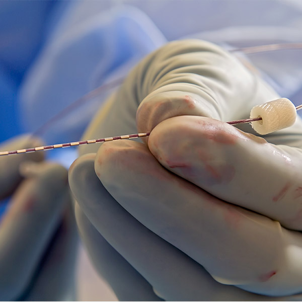
Cardinale outlines the steps involved in the updated workflow: ) Whether performed by the traditional or updated workflow, the goal of SEEG is to provide the surgeon with highly precise information on the location of the epileptogenic zone, used for planning epilepsy surgery. Using the software program from an image-guided neurosurgical robot, the reconstructed data are used to plan the surgical approach, or "trajectory." Robot-assisted surgery was then performed to implant the electrodes. A key part of the development process was creating a "homemade" computer script to automate the necessary series of data processing steps. The digital imaging data then undergo processing for reconstruction, resulting in the creation of a detailed computerized model of the brain and of the vascular tree. Before surgery, the patient undergoes 3-D magnetic resonance imaging and 3-D digital subtraction angiography. Cardinale and colleagues have been working to develop an updated SEEG workflow allowing a one-step surgical technique. The traditional SEEG technique includes two surgical steps: 3-D imaging of the brain blood vessels (stereotactic angiography) followed by electrode implantation. Originally developed by French researchers named Talairach and Bancaud, SEEG uses electrodes implanted in the brain to localize the epileptogenic zone-the area in which seizures originate. The researchers describe the development of and initial experience with an updated SEEG technique for planning epilepsy surgery. Stereo EEG Technique Updated and Simplified Their "updated workflow" combines sophisticated imaging data reconstructions and robot-assisted surgery, "providing essential information in the most complex cases of drug-resistant epilepsy." Francesco Cardinale of Niguarda Ca' Granda Hospital, Milan, and colleagues. "SEEG is a safe and accurate procedure for invasive assessment of the epileptogenic zone," according to the new report by Dr.

The journal is published by Lippincott Williams & Wilkins, a part of Wolters Kluwer Health.

(March 12, 2013) – For patients with "drug-resistant" epilepsy requiring surgery, an updated stereoelectroencephalography (SEEG) technique provides a more efficient process for obtaining critical data for surgical planning, according to a study in the March issue of Neurosurgery, official journal of the Congress of Neurological Surgeons.


 0 kommentar(er)
